Shoulder Tendon Anatomy : Shoulder Joint Anatomy Physiology Movement Exercise. Home » what is frozen shoulder » shoulder anatomy. Shoulder tendon anatomy (page 1). The tendons and the muscles come next. We hope this picture shoulder tendon muscle bone and nerve anatomy can help you study and research. Shoulder joint allows lifting, pushing and pulling by upper extremity.
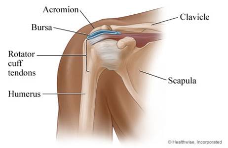
Your shoulder is made up of three bones: Shoulder muscles and shoulder tendons. The subacromial bursa lies on the top portion of the supraspinatus tendon. An image depicting shoulder anatomy can be seen below. Infraspinatus and teres minor tendon.
Shoulder Impingement Boise Rotator Cuff Tendons Boise Eagle Id from www.shouldersurgeon.com Just remember the articulating surfaces. Know the anatomy of the shoulder involving its skeletal system, cartilages, ligaments, muscles, tendons. In this episode of eorthopodtv, orthopaedic surgeon randale c. The scapula (shoulder blade), clavicle (collarbone) and humerus (upper arm bone). The shoulder joint (glenohumeral joint) is a ball and socket joint between the scapula and the humerus. The biceps tendon begins at the top of the shoulder socket (the glenoid) and then passes across the front of the shoulder to connect to the biceps muscle. Upper limb trauma programme injuries. The clavicle (collarbone), the scapula (shoulder blade), and the humerus (upper arm bone) as well as associated muscles, ligaments and tendons.
Muscles allow us to move by pulling on bones.
Shoulder tendonitis is inflammation of your rotator cuff or bicep tendons, often caused by overuse of the arms such as in baseball, weight lifting, and racket sports. Upper limb trauma programme injuries. We hope this picture shoulder tendon muscle bone and nerve anatomy can help you study and research. The human shoulder is made up of three bones: Shoulder tendonitis is the inflammation, irritation and swelling of the tendons in the rotator cuff and bicep. Infraspinatus and teres minor tendon. For more anatomy content please follow us and visit our website: Home » what is frozen shoulder » shoulder anatomy. Sechrest, md narrates an animated tutorial on the basic anatomy of the shoulder. Shoulder muscles and shoulder tendons. An image depicting shoulder anatomy can be seen below. The scapula (shoulder blade), clavicle (collarbone) and humerus (upper arm bone). In this episode of eorthopodtv, orthopaedic surgeon randale c.
In this episode of eorthopodtv, orthopaedic surgeon randale c. Your shoulder is made up of three bones: Muscles allow us to move by pulling on bones. Anatomy of the canine shoulder (scapula, humerus, ligaments, shoulder joint, muscles and tendons) on ct. The shoulder is made up of three bones:
Shoulder Cartilage And Tendon Injuries My Doctor Online from mydoctor.kaiserpermanente.org Sechrest, md narrates an animated tutorial on the basic anatomy of the shoulder. The shoulder is made up of three bones: The biceps tendon begins at the top of the shoulder socket (the glenoid) and then passes across the front of the shoulder to connect to the biceps muscle. Your upper arm bone (humerus), your once the ligaments, tendons, and muscles around the shoulder become loose or torn, dislocations can occur. In addition to shoulder dislocations, other common injuries include rotator cuff tendon tears and broken bones including the humerus and collar terry gc, chopp tm. It is the major joint connecting the upper limb to the trunk. Infraspinatus and teres minor tendon. In this episode of eorthopodtv, orthopaedic surgeon randale c.
The tendons and the muscles come next.
The tendons of the rotator cuff are the next layer in the shoulder joint. An image depicting shoulder anatomy can be seen below. Infraspinatus and teres minor tendon. Shoulder tendonitis is the inflammation, irritation and swelling of the tendons in the rotator cuff and bicep. The subacromial bursa lies on the superior aspect of the supraspinatus tendon (see the images below). The tendons and the muscles come next. The nerves supply all the structures above and make them work. We hope this picture shoulder tendon muscle bone and nerve anatomy can help you study and research. The long head biceps tendon travels through the shoulder joint making it more prone to injury such as a partial tear, rupture. The biceps tendon begins at the top of the shoulder socket (the glenoid) and then passes across the front of the shoulder to connect to the biceps muscle. Just remember the articulating surfaces. You can see these areas marked with an x in the shoulder anatomy diagram above. Your upper arm bone (humerus), your once the ligaments, tendons, and muscles around the shoulder become loose or torn, dislocations can occur.
In addition to shoulder dislocations, other common injuries include rotator cuff tendon tears and broken bones including the humerus and collar terry gc, chopp tm. The shoulder joint is highly mobile and relies on coordination between various muscles, tendons due to its complex anatomy the shoulder is prone to injuries and to degenerative wear and tear such. Upper limb trauma programme injuries. The nerves supply all the structures above and make them work. You can see these areas marked with an x in the shoulder anatomy diagram above.
Shoulder Girdle These Bones Of Mine Shoulder Anatomy Shoulder Joint Anatomy Shoulder Muscles from i.pinimg.com Your shoulder is made up of three bones: Functional anatomy of the shoulder. Shoulder tendonitis is the inflammation, irritation and swelling of the tendons in the rotator cuff and bicep. Anatomy of the canine shoulder (scapula, humerus, ligaments, shoulder joint, muscles and tendons) on ct. Shoulder tendon anatomy (page 1). Shoulder and pectoral region medicine 300 with mustafa/ulasli at gaziantep university. The nerves supply all the structures above and make them work. You can see these areas marked with an x in the shoulder anatomy diagram above.
Related online courses on physioplus.
The tendons of the rotator cuff are the next layer in the shoulder joint. Functional anatomy of the shoulder. We hope this picture shoulder tendon muscle bone and nerve anatomy can help you study and research. It is the major joint connecting the upper limb to the trunk. Shoulder joint allows lifting, pushing and pulling by upper extremity. You can see these areas marked with an x in the shoulder anatomy diagram above. The shoulder joint is highly mobile and relies on coordination between various muscles, tendons due to its complex anatomy the shoulder is prone to injuries and to degenerative wear and tear such. The tendons and the muscles come next. The human shoulder is made up of three bones: Muscles allow us to move by pulling on bones. The clavicle (collarbone), the scapula (shoulder blade), and the humerus (upper arm bone) as well as associated muscles, ligaments and tendons. The shoulder joint is the connection between the chest and the upper extremity. The shoulder joint (glenohumeral joint) is a ball and socket joint between the scapula and the humerus.
Share :
Post a Comment
for "Shoulder Tendon Anatomy : Shoulder Joint Anatomy Physiology Movement Exercise"


Post a Comment for "Shoulder Tendon Anatomy : Shoulder Joint Anatomy Physiology Movement Exercise"